
What Does a Chest X-Ray Show?
Common reasons doctors order chest X-rays, what the images may reveal, and what can happen next.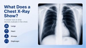
A chest X-ray is one of the most common imaging tests. Your doctor may order one if you have a persistent cough, shortness of breath, chest pain, fever, or another concern involving the lungs or chest. The images can also provide useful information about the heart, ribs, and nearby structures.
A chest X-ray cannot answer every question, but it can often give your healthcare provider a helpful first look at what may be going on.
What Does a Chest X-Ray Look At?
A chest X-ray creates images of several structures in the chest, including:
- The lungs
- The heart
- The ribs
- The diaphragm
- The airways
- Some surrounding soft tissues
Because these structures overlap on a standard X-ray, the radiologist looks at the overall pattern of the image and checks for anything that appears unusual.
Why Might Your Doctor Order One?
A chest X-ray may be recommended for several reasons. Common examples include:
- Ongoing cough
- Shortness of breath
- Chest pain
- Fever with concern for infection
- Follow-up of a known lung condition
- Certain heart-related concerns
- Evaluation after an injury
The reason for the exam depends on your symptoms and what your provider is trying to understand.
Can a Chest X-Ray Show Pneumonia?
Yes, a chest X-ray can sometimes show changes in the lungs that are consistent with pneumonia.
Pneumonia can cause areas of the lung to look different from the surrounding tissue, which may help your doctor support the diagnosis.
That said, not every case of pneumonia appears clearly on an X-ray, especially very early in the illness. Your provider will also consider your symptoms, physical exam, and other test results.
Can It Show Fluid Around the Lungs?
A chest X-ray can sometimes show fluid collecting around the lungs, a condition known as pleural effusion. The amount and location of the fluid can affect how clearly it appears.
If your doctor needs more detail, another imaging test may be recommended.
Can a Chest X-Ray Show Heart Problems?
A chest X-ray can provide some information about the size and shape of the heart.
For example, the heart may appear enlarged in certain conditions. The images can also show some changes in the lungs that may be related to heart problems.
A chest X-ray does not provide a complete picture of how the heart is functioning, so additional tests may be needed depending on the concern.
Can a Chest X-Ray Detect Lung Cancer?
A chest X-ray may sometimes reveal a mass or another abnormal area in the lungs, but it is not a reliable screening test for lung cancer.
Small or subtle cancers may not be visible on a standard chest X-ray.
If a suspicious area is seen, or if your doctor remains concerned despite a normal X-ray, CT imaging may be recommended for a more detailed look.
What Else Can a Chest X-Ray Show?
Depending on the situation, a chest X-ray may also help identify:
- A collapsed lung
- Certain bone injuries
- Changes related to chronic lung disease
- Some infections
- The position of certain medical devices
The images are interpreted in the context of your symptoms and medical history.
What If the Chest X-Ray Is Normal?
A normal chest X-ray can be reassuring, but it does not rule out every possible cause of symptoms. Some conditions are too small, too early, or not well suited to X-ray imaging.
If you continue to have symptoms, your healthcare provider may recommend another test or a different type of evaluation.
What Happens If Something Abnormal Is Found?
An abnormal finding does not automatically mean something serious. Your doctor may recommend:
- Comparing the image with an older X-ray
- Repeating the study later
- Ordering a CT scan
- Performing blood tests
- Referring you to another specialist
The next step depends on what the finding looks like and how it fits with your symptoms.
What Happens During the Exam?
A chest X-ray is usually straightforward. A technologist will position you for the images, and you may be asked to stand, sit, or lie down depending on the exam.
You will likely be asked to take a deep breath and hold it briefly while the image is taken. This helps the lungs expand so they can be seen more clearly.
The X-ray itself does not hurt.
What Happens After the X-Ray?
Once the images are complete, a radiologist reviews them and prepares a report for your referring provider. Your provider will then consider the results along with your symptoms, physical exam, and any other testing.
Depending on the findings, you may need no further imaging, or your doctor may recommend another step.
X-Ray in Denville, NJ
If your healthcare provider recommends a chest X-ray or another diagnostic X-ray, ImageCare Radiology offers X-rays in Denville, NJ.
Chest X-rays can help give your provider a clearer look at the lungs, heart, and surrounding structures when symptoms or other concerns need further evaluation.
A Useful First Look at Chest Symptoms
A chest X-ray can provide valuable information when you are dealing with symptoms such as cough, shortness of breath, chest pain, or fever.
It may help identify changes in the lungs, heart, or nearby structures and give your healthcare provider a clearer idea of what should happen next.
If the images do not fully explain your symptoms, that information can still be useful in deciding whether another test or follow-up is needed.