
Upright MRI for Herniated Discs: What Patients Should Know
Back pain is one of the most common reasons people seek medical care. While many cases improve with rest, physical therapy, or other conservative treatments, persistent symptoms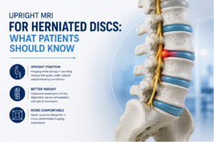 sometimes require a closer look at the spine. One potential cause of ongoing back or neck pain is a herniated disc.
When physicians need detailed images of the spine, MRI is often the imaging study of choice. For some patients, an upright MRI offers an alternative to traditional MRI by allowing imaging to be performed in a weight-bearing or seated position.
If your doctor has recommended imaging for a suspected herniated disc, understanding how upright MRI works and what information it can provide may help you feel more prepared for your exam.
What Is a Herniated Disc?
The spine is made up of a series of bones called vertebrae. Between these vertebrae are soft, cushion-like discs that help absorb shock and support movement.
A herniated disc occurs when part of a disc pushes beyond its normal boundary. In some cases, the displaced disc material may irritate or compress nearby nerves, potentially contributing to symptoms such as:
- Back pain
- Neck pain
- Pain radiating into the arms or legs
- Numbness or tingling
- Muscle weakness
Symptoms and severity vary from person to person, which is why imaging is often an important part of the evaluation process.
Why MRI Is Commonly Used for Herniated Discs
Unlike X-rays, which primarily show bones, MRI provides detailed images of soft tissues. This allows physicians to evaluate:
- Intervertebral discs
- Spinal nerves
- The spinal cord
- Ligaments
- Surrounding soft tissues
MRI can help identify the location and extent of a herniated disc while providing important information about nearby structures.
What Is an Upright MRI?
An upright MRI, sometimes called a stand-up MRI, is designed to allow imaging while a patient is sitting, standing, or positioned more naturally than in a traditional lying-down MRI.
This approach may provide additional perspectives because the spine is evaluated while bearing weight rather than only while lying flat.
Depending on the clinical situation, physicians may find this information helpful when evaluating certain spinal conditions.
Why Weight-Bearing Imaging Matters
Many patients notice that their symptoms worsen when standing, walking, sitting for extended periods, or performing daily activities.
However, traditional MRI imaging is performed while lying down, which may reduce some of the natural forces acting on the spine.
An upright MRI allows physicians to observe spinal structures under conditions that more closely resemble normal daily posture.
This can provide additional information about:
- Disc alignment
- Spinal positioning
- Nerve compression
- Changes that occur during weight-bearing
The usefulness of weight-bearing imaging depends on each patient’s individual condition and symptoms.
Potential Benefits for Some Patients
A More Comfortable Experience
Many patients appreciate the more open design of an upright MRI system. For individuals who experience anxiety in enclosed spaces, the open configuration may feel more comfortable than a traditional MRI scanner.
Imaging in Functional Positions
In some cases, symptoms occur primarily while sitting, standing, or maintaining certain postures. An upright MRI may allow physicians to evaluate the spine in positions that better reflect how symptoms occur during daily activities.
Evaluation of Complex Spine Conditions
In addition to herniated discs, upright MRI may sometimes be used when evaluating:
- Degenerative disc disease
- Spinal stenosis
- Certain alignment concerns
- Chronic neck pain
- Chronic back pain
The specific imaging approach depends on the physician’s clinical goals and the patient’s symptoms.
What Happens During an Upright MRI?
The procedure is similar to a traditional MRI in many respects. During the exam:
- A technologist positions you appropriately.
- Images are obtained using magnetic fields and radio waves.
- You will need to remain still during image acquisition.
- Multiple views of the spine may be captured.
The exam is noninvasive and does not use ionizing radiation. The length of the study varies depending on the area being examined.
Can Upright MRI Diagnose a Herniated Disc?
An MRI can provide detailed images that help physicians evaluate spinal discs and surrounding structures. However, imaging findings are only one part of the diagnostic process.
Physicians also consider:
- Symptoms
- Medical history
- Physical examination findings
- Additional testing when appropriate
A herniated disc seen on imaging does not always cause symptoms, and symptoms do not always correlate perfectly with imaging findings. This is why MRI results are interpreted within the context of the patient’s overall clinical picture.
Who May Benefit From an Upright MRI?
An upright MRI may be considered for patients who:
- Have symptoms that worsen while standing or sitting
- Experience discomfort lying flat
- Prefer a more open MRI environment
- Need additional spinal evaluation based on physician recommendations
Your healthcare provider can determine whether an upright MRI is appropriate for your situation.
Upright MRI in Brunswick, NJ
At ImageCare Radiology, patients seeking an upright MRI in Brunswick, NJ, have access to advanced weight-bearing MRI technology designed to provide detailed spinal imaging in a more open and comfortable environment. These studies can help physicians evaluate herniated discs and other spinal conditions while gathering important information to support diagnosis and treatment planning.
Getting a Clearer Picture of Spine Health
When back or neck pain persists, diagnostic imaging can play an important role in understanding what may be contributing to symptoms. For some patients with suspected herniated discs, an upright MRI offers a unique way to evaluate the spine in a weight-bearing position while providing the detailed soft-tissue images MRI is known for.
By helping physicians better assess discs, nerves, and spinal structures, upright MRI can provide valuable information that supports informed treatment decisions and a more personalized approach to care.