
Understanding Cross-Sectional Imaging: How CT Technology Works
Medical imaging has come a long way from the days when physicians relied solely on traditional X-rays to evaluate injuries and illnesses. Today, advanced imaging technologies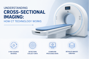 provide far more detailed views of the body’s internal structures, helping healthcare providers make informed diagnostic and treatment decisions.
One of the most widely used imaging tools is the CT scan, which uses cross-sectional imaging to create highly detailed pictures of organs, bones, blood vessels, and soft tissues.
But what exactly does “cross-sectional imaging” mean, and how does CT technology work?
Understanding the basics of CT imaging can help patients feel more comfortable and informed when preparing for an exam.
What Is Cross-Sectional Imaging?
Cross-sectional imaging refers to the creation of detailed “slice-like” images of the body.
Instead of producing a single flat image like a traditional X-ray, cross-sectional imaging allows physicians to view the body layer by layer.
You can think of it as looking at individual pages within a book rather than only seeing the cover.
These detailed slices help healthcare providers examine structures from multiple angles and better understand what may be happening inside the body.
What Is a CT Scan?
CT stands for computed tomography.
A CT scan combines X-ray technology with sophisticated computer processing to generate detailed cross-sectional images.
During the exam, the CT scanner captures numerous images from different angles around the body. A computer then assembles these images into detailed slices that can be reviewed individually or reconstructed into three-dimensional views.
This level of detail allows physicians to evaluate many structures that may be difficult to assess with conventional X-rays alone.
How CT Technology Captures Images
A CT scanner consists of a large circular opening, often described as doughnut-shaped. During the exam:
- The patient lies on a motorized table.
- The table moves slowly through the scanner.
- An X-ray source rotates around the body.
- Detectors collect imaging data from multiple angles.
- A computer processes the information into cross-sectional images.
The process is fast, precise, and designed to capture highly detailed diagnostic information.
Why Cross-Sectional Images Are Valuable
Traditional X-rays can provide important information, but structures may overlap within a single image. Cross-sectional imaging helps reduce this limitation by separating anatomy into individual slices.
This allows physicians to visualize:
- Internal organs
- Bones and joints
- Blood vessels
- Soft tissues
- Areas of injury or disease
The ability to examine structures layer by layer often provides greater clarity and diagnostic confidence.
Creating Three-Dimensional Views
One of the remarkable advantages of modern CT technology is its ability to create three-dimensional reconstructions.
Using the cross-sectional images, advanced computer software can generate 3D representations of specific anatomical structures.
These views may help physicians:
- Better understand complex anatomy
- Evaluate certain injuries
- Plan procedures
- Assess the relationship between structures
The original cross-sectional images remain the foundation of the exam, while 3D reconstructions provide additional perspectives when needed.
What Conditions Can CT Scans Help Evaluate?
Because CT imaging provides detailed views of many parts of the body, it is used in a wide range of clinical situations.
Physicians may recommend CT scans to help evaluate:
- Injuries and trauma
- Abdominal pain
- Chest conditions
- Certain cancers
- Blood vessel abnormalities
- Neurological concerns
- Unexplained symptoms
The specific role of CT imaging depends on the patient’s symptoms, medical history, and the clinical question being investigated.
How CT Differs From a Traditional X-Ray
Although CT scans use X-rays, they provide significantly more information than a standard radiograph.
- Traditional X-Ray
- Produces a single image
- Useful for evaluating many bone-related conditions
- Often serves as an initial imaging study
CT Scan
- Produces multiple cross-sectional images
- Provides greater anatomical detail
- Allows viewing from multiple perspectives
- Can generate 3D reconstructions
Both imaging modalities are valuable, but they serve different purposes.
Does Every CT Scan Use Contrast?
Not all CT scans require contrast material.
When needed, contrast can help certain tissues, organs, and blood vessels appear more clearly on imaging. Depending on the examination, contrast may be:
- Administered through an IV
- Swallowed as a liquid
- Used in another exam-specific manner
Your healthcare provider and imaging team will determine whether contrast is appropriate for your study.
What Happens During the Exam?
For most patients, the CT scan itself is relatively quick and straightforward. During the procedure:
- You will lie on the examination table.
- The table moves through the scanner.
- The scanner rotates around the area being imaged.
- Images are captured from multiple angles.
- You may be asked to hold your breath briefly during certain portions of the scan.
The exam is painless, and many studies are completed within minutes.
How Radiologists Interpret CT Images
Once the scan is complete, a radiologist reviews the images. Radiologists are physicians with specialized training in medical imaging interpretation.
They evaluate:
- Anatomical structures
- Areas of concern
- Potential abnormalities
- Findings relevant to the physician’s clinical question
A report is then sent to the referring provider, who discusses the results and any recommended next steps with the patient.
CT Scan in Hackettstown, NJ
At ImageCare Radiology, patients seeking a CT scan in Hackettstown, NJ, have access to advanced imaging technology designed to produce detailed cross-sectional images that support accurate diagnoses and informed treatment planning.
Experienced radiologists carefully interpret each study to provide valuable information for patients and their healthcare providers.
Looking Beneath the Surface
Cross-sectional imaging has transformed the way physicians evaluate injuries, illnesses, and medical conditions. By creating detailed slice-by-slice views of the body, CT technology provides information that can often reveal details not visible with conventional imaging alone.
Whether investigating symptoms, assessing an injury, or monitoring a medical condition, CT scans remain one of the most powerful diagnostic tools available, helping healthcare providers gain a clearer understanding of what is happening beneath the surface.