
How Digital X-Rays Improve the Imaging Experience
What modern digital X-ray technology means for image review, sharing, and your visit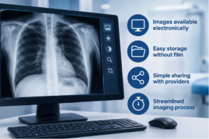
If you have had an X-ray in the last several years, there is a good chance it was digital.
The exam itself may not feel dramatically different from older X-rays, but the way the images are captured, reviewed, stored, and shared has changed quite a bit.
Digital X-ray technology allows images to appear electronically instead of being developed on physical film. For patients, that can make the overall imaging process more efficient and easier to manage from start to finish.
What Is a Digital X-Ray?
A digital X-ray uses the same basic X-ray technology doctors have relied on for years, but the image is captured electronically.
Instead of exposing film that later has to be developed, the image is available on a computer for the technologist and radiologist to review.
That means there is no physical film to process, store, or transport.
How Is That Different From Traditional Film X-Ray?
Traditional X-rays were recorded on physical film, much like photographs.
The film had to be developed before anyone could review the image. If the image was not clear enough, the problem might not be obvious until after processing.
Digital imaging removes that step.
Once the image is taken, it can be viewed electronically, which helps the technologist confirm that the necessary views were captured.
What Does Digital Imaging Change for Patients?
The biggest changes usually happen behind the scenes. Digital X-rays can make it easier to:
- Review images soon after they are taken
- Store images electronically
- Compare newer images with older studies
- Share images with referring providers
- Avoid handling or transporting physical film
For most patients, the exam still feels familiar. A technologist positions the part of the body being examined, asks you to remain still briefly, and takes the needed images.
Can the Images Be Reviewed More Easily?
Yes. Digital X-ray images can be viewed on a computer, which gives the radiologist more flexibility when examining them.
Certain viewing settings, such as brightness or contrast, can be adjusted to make specific details easier to assess.
That does not change what was captured in the original image. It simply gives the radiologist more options for viewing the information that is already there.
Does Digital X-Ray Mean Faster Results?
Digital technology makes the images available more quickly for review, but that does not always mean you will receive your final results immediately.
A radiologist still needs to examine the images and prepare a report. Your referring provider then reviews that report and discusses the findings with you.
The electronic format helps streamline the process, but professional interpretation still takes time.
Are Digital X-Rays More Accurate?
Digital technology offers practical advantages, but image quality still depends on proper positioning, appropriate technique, and careful interpretation.
The technology can make images easier to review and manage, but it does not replace the skill of the technologist who captures them or the radiologist who reads them.
A clear, useful X-ray still starts with getting the right images.
Do Digital X-Rays Still Use Radiation?
Yes. Digital X-rays still use ionizing radiation.
The difference is how the image is recorded, not the type of energy used to create it.
When an X-ray is medically appropriate, the goal is to obtain the information your healthcare provider needs while avoiding unnecessary exposure.
If you are pregnant or think you may be pregnant, tell your healthcare provider or technologist before the exam.
What Can Digital X-Rays Be Used For?
Digital X-rays can be used for many different types of exams, including imaging of the:
- Chest
- Spine
- Arms and legs
- Hands and feet
- Knees and other joints
- Abdomen
- Bones after an injury
The type of X-ray your doctor orders depends on your symptoms and the area that needs to be examined.
What Happens During a Digital X-Ray?
The process is usually simple.
A technologist will position you for the images your doctor requested. Depending on the exam, you may stand, sit, or lie down.
You may need to change position so the area can be viewed from more than one angle.
The X-ray itself does not hurt, although positioning can be uncomfortable if you are already injured or sore. Let the technologist know if a movement is difficult.
What Happens After the Images Are Taken?
Once the exam is complete, a radiologist reviews the images and prepares a report for your referring provider.
Because the images are stored electronically, they can also be compared with previous studies when those are available.
Your provider then uses the report along with your symptoms, physical exam, and other information to decide what happens next.
X-Rays in Millburn, NJ
ImageCare Radiology offers X-rays in Millburn, NJ, using digital radiography technology.
Digital imaging allows X-ray images to be captured, stored, and shared electronically, helping referring providers access the information they need to evaluate injuries, joint concerns, chest symptoms, and other medical issues.
A More Streamlined Way to Handle X-Ray Images
Digital X-ray technology has changed how images move through the healthcare system.
There is no film to develop, no physical image to carry from one office to another, and images can be reviewed and stored electronically.
For patients, the exam itself remains simple and familiar, while the technology behind it helps make the rest of the imaging process easier to manage.