
Digital X-Ray vs. Film X-Ray: What’s the Difference?
If you have had an X-ray in recent years, there is a good chance it was digital.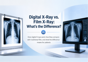
That does not mean the basic purpose of the exam has changed. X-rays are still used to help doctors look at bones, joints, the chest, and other parts of the body. The main difference is how the image is captured and viewed.
Traditional film X-rays had to be processed much like photographic film. Digital X-rays create an electronic image that can be viewed on a computer shortly after it is taken.
For patients, that usually means a smoother and more efficient imaging process.
What Is a Digital X-Ray?
A digital X-ray uses X-rays to create an image, but instead of recording that image on film, it is captured electronically.
Once the image is taken, the technologist can review it on a computer screen. The radiologist can then examine the image in detail and prepare a report for your referring provider. Because the image is digital, it can also be stored and shared electronically.
How Did Film X-Rays Work?
Before digital imaging became common, X-ray images were recorded on physical film.
That film had to be developed before the image could be reviewed. If the image was not clear enough, another image might need to be taken after the film was processed.
Film also had to be stored physically and transported if another provider needed to review it. Digital imaging removed many of those extra steps.
What Is the Biggest Difference for Patients?
For most patients, the actual exam feels very similar. You are still positioned by a technologist, asked to stay still for a moment, and the image is taken quickly.
The main difference happens after the image is captured.
With digital X-rays, the technologist can see the image sooner and confirm whether the necessary views were obtained. The images can also be sent electronically for review rather than relying on physical film.
That can make the overall process more efficient.
Are Digital X-Rays More Accurate?
Digital technology offers several advantages, but it is important not to think of it as automatically producing a “better diagnosis.”
The quality of any X-ray still depends on several things, including:
- Proper positioning
- The correct imaging technique
- The area being examined
- The quality of the equipment
- Professional interpretation by a radiologist
One advantage of digital imaging is that the images can be adjusted on a screen to help the radiologist review certain details more clearly.
But the technology itself does not replace the experience and judgment involved in interpreting the exam.
Do Digital X-Rays Still Use Radiation?
Yes. Digital X-rays still use ionizing radiation to create images.
The difference is how the image is recorded, not the fact that X-rays are being used. When an X-ray is medically appropriate, the goal is to use the amount of radiation needed to obtain a useful diagnostic image while avoiding unnecessary exposure.
If you are pregnant or think you may be pregnant, tell your healthcare provider or technologist before the exam.
Why Did Digital X-Rays Replace Film in Many Settings?
Digital imaging offers practical advantages for both patients and healthcare providers. Images can be:
- Viewed quickly
- Stored electronically
- Shared more easily with other providers
- Reviewed on a computer without needing physical film
This is especially helpful when more than one provider is involved in your care or when previous images need to be compared with newer ones.
Digital storage also reduces the need to keep large amounts of physical film.
Can Digital X-Rays Be Enhanced?
Yes, within limits. Radiologists can adjust certain viewing settings on digital images, such as brightness or contrast, to help examine different details.
That does not mean the image can be changed to reveal something that was never captured. Good positioning and proper technique still matter from the start.
Think of it more like having better tools for viewing the image rather than changing what the X-ray actually showed.
What Can Digital X-Rays Be Used For?
Digital X-rays can be used for many of the same reasons traditional film X-rays were used. Common examples include evaluating:
- Suspected fractures
- Joint injuries
- Arthritis-related changes
- Chest symptoms
- Spine concerns
- Certain abdominal conditions
- Bone alignment
The type of X-ray your doctor orders depends on your symptoms and the part of the body that needs to be examined.
What Happens After a Digital X-Ray?
Once the images are complete, a radiologist reviews them and prepares a report for your referring provider. Your provider then considers the X-ray findings together with your symptoms, physical exam, and any other test results.
Depending on what the images show, the next step may be treatment, follow-up, or another type of imaging.
X-Rays in Morristown, NJ
If your healthcare provider recommends diagnostic imaging, ImageCare Radiology offers X-Rays in Morristown, NJ, using modern digital imaging technology.
Digital X-rays allow images to be captured and reviewed efficiently while giving your referring provider the information needed to better understand injuries, joint concerns, chest symptoms, and other medical issues.
The Technology Changed, but the Goal Stayed the Same
Digital X-rays have made medical imaging faster and easier to manage, especially when it comes to viewing, storing, and sharing images.
But the purpose of the exam is still simple: to give your healthcare provider a clearer look at what may be happening inside your body.
For patients, that means the benefits of modern technology without a dramatically different exam experience.