
Breast Ultrasound for a Lump: What Can It Show?
How ultrasound helps radiologists evaluate a breast lump and determine what may come next.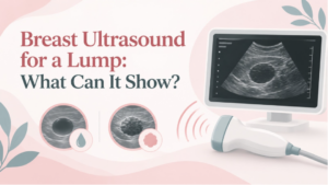
Finding a breast lump can raise a lot of questions. Many lumps are benign, but a new or changing lump should still be evaluated so your healthcare provider can better understand what is causing it.
Breast ultrasound is one of the imaging tools that may be used during that process. It gives the radiologist a focused look at the area of concern and can reveal details that are difficult to determine through touch alone.
Why Might a Breast Lump Need an Ultrasound?
Sometimes a lump is something you notice yourself. Other times, a healthcare provider may feel an area during an exam, or another breast imaging study may show something that deserves a closer look.
Ultrasound uses sound waves to create images of the tissue beneath the skin. It can be directed right at the area of concern, helping the radiologist see what the lump looks like and how it relates to the surrounding breast tissue.
Which imaging test comes first depends on factors such as age and the reason for the exam. For some patients, ultrasound is the first step. For others, diagnostic mammography and ultrasound are used together.
Can Ultrasound Tell Whether a Lump Is a Cyst or a Solid Mass?
Often, yes. This is one of the main reasons ultrasound is useful when evaluating a breast lump.
A simple cyst is filled with fluid, while a solid mass is made of tissue. Ultrasound can often distinguish between the two and can also show findings that contain both cystic and solid components.
A solid finding does not mean breast cancer. Many benign conditions can create solid breast masses. What matters is how the finding looks as a whole and whether its imaging features suggest that anything further needs to be done.
The RadiologyInfo guide to breast ultrasound explains how ultrasound is used to characterize breast abnormalities found during an exam, mammogram, or MRI.
What Does the Radiologist Look For?
The radiologist looks at several characteristics of the area, including its shape, margins, internal appearance, and relationship to nearby tissue.
Those features help the radiologist classify the finding and recommend an appropriate next step. Breast imaging reports use the standardized BI-RADS system to communicate whether a finding is negative, benign, probably benign, suspicious, or requires another type of evaluation.
That assessment comes from the entire imaging picture. It is not something that can reliably be determined from one ultrasound feature alone.
Can Breast Ultrasound Diagnose Cancer?
Ultrasound can show whether a finding has features that are more or less concerning, but it cannot always determine exactly what a solid mass is.
If the imaging looks suspicious, the radiologist may recommend a biopsy. A small tissue sample is then examined by a pathologist to determine what the cells or tissue actually represent. Ultrasound itself does not provide that tissue diagnosis.
Many findings do not lead to biopsy. Some can be confidently identified as benign, while others may be followed with additional imaging to make sure they remain stable.
Will I Also Need a Mammogram?
You might. Breast ultrasound and mammography provide different information, so they are often used together rather than treated as interchangeable tests.
For a new palpable lump, ultrasound is usually the first imaging test in patients younger than 30. Beginning at age 30, diagnostic mammography or 3D mammography and ultrasound commonly play complementary roles. Your provider and radiologist will determine which imaging is appropriate for your situation.
Breast ultrasound also does not replace mammography when mammography is recommended for routine breast cancer screening. Some cancers, particularly those that appear as calcifications, may be visible on mammography but not ultrasound.
What Happens After a Breast Ultrasound?
The answer depends on what the radiologist sees and how well the imaging matches the area you or your healthcare provider can feel.
The result may show a benign cyst or another reassuring finding. In other cases, the radiologist may recommend follow-up imaging, another diagnostic test, or a biopsy.
It is also possible for imaging to look normal even when a lump can still be felt. If a new lump persists or remains clinically concerning, follow up with your healthcare provider rather than assuming that normal imaging means no further evaluation is needed.
Ultrasound in Paterson, NJ
PINK Breast Center provides ultrasound in Paterson, NJ, as part of our breast imaging services. The Paterson center specifically uses breast ultrasound to evaluate lumps and other areas of concern and also offers mammography and breast biopsy services when those tests or procedures are appropriate.
The type of imaging you need will depend on your symptoms, age, previous studies, and the recommendation from your healthcare provider.
When a Breast Lump Needs a Closer Look
A breast lump does not automatically point to cancer. There are many benign reasons for changes in breast tissue, and ultrasound can help narrow down what may be going on.
By showing whether an area is fluid-filled or solid and providing a closer look at its features, breast ultrasound gives the radiologist information that can help guide the next step. Sometimes that means no additional imaging is needed. Sometimes it means follow-up or biopsy. The important part is having a new or changing lump properly evaluated rather than trying to interpret it on your own.