At PINK Breast Center, part of ImageCare Radiology, we provide expert breast imaging including 2D mammograms in Paterson, NJ, in a supportive and respectful environment. Our team understands that breast health appointments can feel stressful, which is why we focus on clear communication, comfort, and compassionate care.
Whether you are visiting for routine mammogram screening or a follow-up diagnostic mammogram, our experienced technologists and radiologists deliver accurate diagnostic imaging services designed to support early detection and informed care decisions. For patients who may benefit from additional imaging, we also offer 3D mammogram technology at our center.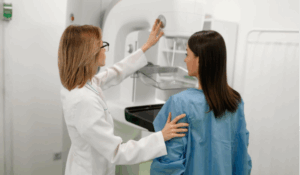
What Is a 2D Mammogram?
A 2D mammogram is a digital X-ray of the breast that allows radiologists to examine internal breast tissue for signs of abnormalities. This form of diagnostic imaging services helps detect early signs of breast cancer, often before symptoms appear.
During the exam, each breast is gently compressed between two plates to spread the tissue evenly. This allows the imaging system to capture clear pictures of the breast structure. While compression may feel slightly uncomfortable for a few seconds, the process is brief and plays an important role in producing accurate images.
A 2D mammogram can help identify:
- Breast masses or lumps
- Areas that may require additional imaging
- Microcalcifications (tiny calcium deposits)
- Structural changes within breast tissue
Regular mammogram screenings are one of the most effective tools for detecting breast cancer early, when treatment is often most successful.
Screening Mammogram vs. Diagnostic Mammogram
At PINK Breast Center, we offer both screening and diagnostic mammogram exams as part of our comprehensive diagnostic imaging services.
Screening Mammogram
A screening exam is recommended for women who have no current symptoms. Annual mammogram screening beginning around age 40 helps detect changes in breast tissue at an early stage.
Diagnostic Mammogram
A diagnostic mammogram is used when additional imaging is needed. This may be recommended if:
- A lump or unusual change is found in the breast
- A previous mammogram showed an area that needs closer evaluation
- There are symptoms such as nipple discharge, pain, or skin changes
Diagnostic exams often include additional images that allow radiologists to evaluate specific areas more closely.
Breast Imaging Designed Around Patient Comfort
At our Paterson location, the PINK Breast Center focuses on providing breast imaging in a calm and supportive setting. Our team understands that every patient’s experience is unique, and we work to make each visit as comfortable and reassuring as possible.
Patients choose our center because we offer:
- Experienced technologists specializing in breast diagnostic imaging services
- Advanced digital mammography equipment
- A patient-focused environment designed for privacy and comfort
- Clear communication before, during, and after your exam
- Efficient scheduling and timely results
For patients who may benefit from additional imaging detail, our center also offers 3D mammogram technology.
When Should You Schedule a Mammogram?
Most women are encouraged to begin regular mammogram screening around age 40 and continue annually or as recommended by their healthcare provider.
Your doctor may suggest earlier screening if you have:
- A personal or family history of breast cancer
- Genetic risk factors
- Previous abnormal breast imaging results
It is also important to contact your provider if you notice changes such as a lump, swelling, nipple discharge, or changes in breast shape or skin texture. These symptoms may require a diagnostic mammogram or additional imaging.
What Happens After Your Mammogram?
After your exam, a board-certified radiologist will review the images from your 2D mammogram.
In many cases, results show normal breast tissue. If an area requires further evaluation, your doctor may recommend additional imaging or follow-up testing. This does not necessarily mean cancer is present, but careful review helps ensure accurate diagnosis and appropriate care.
Your referring provider will discuss your results and any next steps with you.
Schedule Your 2D Mammogram in Paterson, NJ
If you are due for a 2D mammogram, the team at PINK Breast Center is here to help. Our experienced staff provides compassionate care and reliable diagnostic imaging services focused on early detection and patient comfort.
Call today or request your appointment online to schedule your mammogram screening.
Frequently Asked Questions About 2D Mammogram
What is the difference between a screening mammogram and a diagnostic mammogram?
A mammogram screening is a routine exam performed when there are no symptoms. A diagnostic mammogram is used when additional images are needed to evaluate symptoms or investigate an area seen on a previous scan.
How long does a 2D mammogram take?
A typical 2D mammogram takes about 10–15 minutes. The imaging portion itself is very quick, with compression lasting only a few seconds for each image.
Is a mammogram painful?
Most patients feel brief pressure during the exam due to breast compression, but the discomfort is temporary and helps ensure clear imaging results.
How often should I have a mammogram?
Most women should begin annual mammogram screening around age 40, though your physician may recommend a different schedule depending on your personal risk factors.
Do you offer 3D mammograms?
Yes. In addition to 2D mammography, our center also offers 3D mammogram imaging, which can provide additional detail for certain patients.
What if something unusual appears on my mammogram?
If an area looks unusual, your doctor may recommend additional diagnostic imaging services, such as a diagnostic mammogram, ultrasound, or other tests. Many findings turn out to be benign, but follow-up imaging helps ensure an accurate evaluation.