
Breast Ultrasound vs. Mammogram: Understanding the Differences
When it comes to breast health, patients are often familiar with mammograms but may be less certain about the role of breast ultrasound. If your healthcare provider recommends one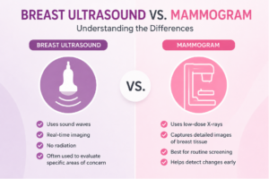 of these exams, or perhaps both, it’s natural to wonder how they differ and why each may be used.
Although breast ultrasound and mammography are both valuable imaging tools, they serve different purposes. Rather than competing technologies, they are often complementary studies that provide different types of information about breast tissue.
Understanding the differences can help patients feel more informed and confident when discussing breast imaging recommendations with their healthcare providers.
What Is a Mammogram?
A mammogram is a specialized X-ray of the breast that is primarily used to screen for and evaluate breast abnormalities.
Mammography can help identify changes within breast tissue that may not be visible or palpable during a physical examination.
Mammograms are commonly used for:
- Routine breast cancer screening
- Evaluating breast symptoms
- Investigating abnormal findings
- Monitoring certain breast conditions
- Follow-up imaging when needed
Because mammography can detect some abnormalities before symptoms develop, it remains an important component of breast health screening.
What Is a Breast Ultrasound?
A breast ultrasound uses sound waves to create images of breast tissue. Unlike mammography, ultrasound does not use radiation. Instead, a handheld transducer sends sound waves into the breast and converts the returning echoes into real-time images.
Breast ultrasound is often used to:
- Evaluate a specific area of concern
- Examine breast lumps
- Further assess abnormalities seen on mammograms
- Help distinguish between certain types of breast findings
- Guide some breast procedures
Since ultrasound produces real-time images, it can provide valuable information about the characteristics of breast tissue and certain breast abnormalities.
How the Technologies Differ
The primary difference is the imaging technology used.
Mammography Uses:
- Low-dose X-rays
- Specialized breast imaging equipment
- Images designed to evaluate breast tissue patterns and abnormalities
Breast Ultrasound Uses:
- Sound waves
- A handheld transducer
- Real-time imaging technology
Each modality reveals different information, which is one reason they are frequently used together rather than as substitutes.
When Mammograms Are Typically Recommended
Mammography is considered the standard imaging tool for routine breast cancer screening. Your healthcare provider may recommend a mammogram for:
- Routine screening based on age and risk factors
- Follow-up evaluation of symptoms
- Investigation of breast changes
- Monitoring previously identified findings
Screening recommendations vary based on individual risk factors, family history, and clinical circumstances.
When Breast Ultrasound May Be Recommended
Breast ultrasound is often used as a diagnostic tool rather than a primary screening exam for most patients. A physician may recommend breast ultrasound when:
- A lump is felt during a physical examination
- An area of concern is identified on a mammogram
- Additional information is needed about a breast abnormality
- Certain breast symptoms require further evaluation
Ultrasound can provide additional detail that helps physicians better understand specific findings within the breast.
Why Dense Breast Tissue Matters
One situation where breast ultrasound may play an important role is in patients with dense breast tissue. Dense breast tissue is common and simply refers to the proportion of fibrous and glandular tissue compared to fatty tissue within the breast.
Dense tissue sometimes makes interpretation more challenging on mammography alone, so healthcare providers may recommend additional imaging in certain situations.
The appropriate imaging approach varies based on each patient’s individual circumstances and clinical needs.
Can a Breast Ultrasound Replace a Mammogram?
This is one of the most common questions patients ask. In most situations, breast ultrasound is not considered a replacement for routine mammographic screening.
The two exams provide different information:
- Mammography is often used for screening and identifying certain breast changes.
- Ultrasound is frequently used to further evaluate specific findings.
Depending on the clinical situation, one study may be recommended, or both may be used together.
What Happens During a Mammogram?
During a mammogram:
- The breast is positioned on specialized imaging equipment.
- Gentle compression is applied briefly to obtain clear images.
- Multiple images are captured from different angles.
- A radiologist reviews the images.
The procedure typically takes only a short time to complete.
What Happens During a Breast Ultrasound?
During a breast ultrasound:
- A technologist applies a small amount of gel to the skin.
- A transducer is moved over the breast.
- Real-time images appear on a monitor.
- Images are reviewed by a radiologist.
The procedure is noninvasive and generally comfortable for most patients.
Why Physicians Often Use Both
Breast imaging is rarely a one-size-fits-all process. Mammography and breast ultrasound each provide unique information that can help physicians evaluate breast tissue more thoroughly.
Together, they may help:
- Clarify findings
- Characterize abnormalities
- Support diagnostic decision-making
- Guide follow-up recommendations
The goal is always to obtain the most accurate information possible while tailoring imaging to each patient’s needs.
Breast Ultrasound in Paterson, NJ
At ImageCare Radiology’s Pink Breast Center, patients seeking a breast ultrasound in Paterson, NJ, have access to advanced breast imaging technology and experienced radiologists dedicated to providing comprehensive breast health evaluations. Breast ultrasound may be used alongside mammography to provide additional information when clinically appropriate.
Understanding the Role of Each Exam
Breast ultrasound and mammography are both important tools in modern breast imaging, but they serve different purposes. Mammograms remain a cornerstone of breast cancer screening, while breast ultrasound often provides additional insight when evaluating specific findings or concerns.
By understanding how these imaging studies differ and how they work together, patients can have more informed conversations with their healthcare providers and feel more confident in their breast health decisions.