
The Role of X-Rays in Orthopedic Injury Diagnosis
Orthopedic injuries can happen suddenly or develop gradually over time. A fall on the ice, a sports injury, an awkward step off a curb, or even repetitive strain can all affect the bones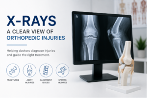 and joints that keep the body moving.
When pain, swelling, or limited mobility occurs, one of the first tools physicians often use is an X-ray. X-rays provide quick, detailed images of bones and joint structures, helping doctors better understand what may be causing symptoms and how an injury should be treated.
For many patients, X-rays are the starting point that helps turn uncertainty into answers.
What Is an Orthopedic Injury?
Orthopedic injuries involve the musculoskeletal system, including:
- Bones
- Joints
- Ligaments
- Tendons
- Muscles
These injuries can range from minor sprains to more serious fractures or joint damage.
Common orthopedic injuries include:
- Broken bones
- Joint dislocations
- Sports injuries
- Stress fractures
- Arthritis-related joint pain
Because symptoms can overlap, imaging is often needed to determine the extent of the injury.
Why X-Rays Are Often the First Imaging Step
X-rays are commonly used as an initial imaging test because they are fast, effective, and particularly useful for evaluating bones and joint alignment.
Physicians often begin with X-rays because they can quickly help identify:
- Fractures
- Dislocations
- Bone alignment problems
- Degenerative joint changes
In many situations, X-rays provide enough information to guide immediate treatment decisions.
What X-Rays Can Detect
Although X-rays are most commonly associated with broken bones, they can reveal much more than fractures alone.
Fractures and Broken Bones
X-rays are especially effective at identifying fractures, including:
- Simple fractures
- Stress fractures
- Displaced fractures
- Hairline breaks
Imaging helps physicians understand the location and severity of the injury.
Joint Dislocations
When a joint moves out of its normal position, X-rays can help confirm the dislocation and assess alignment after treatment.
Common areas evaluated include the:
- Shoulder
- Elbow
- Knee
- Fingers
Arthritis and Degenerative Changes
X-rays may also show signs of wear and tear within joints, including narrowing of joint spaces or bone changes associated with arthritis.
This can help physicians evaluate chronic pain or stiffness.
Bone Alignment and Structural Changes
In some cases, X-rays help identify structural abnormalities or alignment concerns that may contribute to pain or reduced mobility.
When Additional Imaging May Be Needed
While X-rays are extremely useful, they do have limitations. They are best at showing bones and joint alignment, but soft tissue injuries are often more difficult to evaluate.
Depending on the injury, a physician may recommend additional imaging, such as:
- MRI for ligaments, tendons, or cartilage
- CT scans for complex fractures
- Ultrasound for certain soft tissue concerns
Additional imaging does not mean the X-ray was unsuccessful. It simply means more detailed information may be needed.
What to Expect During an Orthopedic X-Ray
Orthopedic X-rays are straightforward and typically take only a few minutes.
During the exam:
- A technologist positions the injured area
- Images are captured from one or more angles
- Patients may be asked to hold still briefly during imaging
The process is noninvasive, and most patients can return to normal activities immediately afterward unless the injury itself limits movement.
Why Timely Imaging Matters
After an injury, some people wait to seek evaluation because they hope symptoms will improve on their own. While minor injuries sometimes do heal with rest, other conditions may worsen without proper diagnosis and treatment.
Prompt imaging can help:
- Confirm or rule out fractures
- Prevent untreated injuries from becoming more serious
- Support faster treatment decisions
- Reduce uncertainty about symptoms
Even injuries that initially seem minor can occasionally involve underlying damage that is not obvious externally.
X-Rays in Rockaway, NJ
At ImageCare Radiology, patients seeking X-rays in Rockaway, NJ, have access to digital imaging designed to provide fast, detailed orthopedic evaluation. Experienced technologists and radiologists work closely with referring providers to help ensure accurate imaging and timely results.
Whether evaluating a sports injury, fall, or ongoing joint pain, high-quality imaging helps physicians make more informed care decisions.
A Key Part of Orthopedic Care
X-rays remain one of the most important tools in orthopedic diagnosis because they provide quick insight into how bones and joints are functioning after injury.
While additional imaging is sometimes needed, X-rays are often the first step toward understanding the problem, guiding treatment, and helping patients begin the recovery process with greater clarity and confidence.