
How Chest X-Rays Help Evaluate Lung Conditions
When someone develops a persistent cough, shortness of breath, chest discomfort, or other respiratory symptoms, physicians often need a way to look beyond what can be observed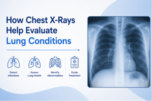 during a physical examination. One of the most common diagnostic tools used in these situations is a chest X-ray.
Chest X-rays provide a quick and noninvasive way to evaluate the lungs and surrounding structures within the chest. While they do not diagnose every lung condition on their own, they often provide important information that helps physicians determine what may be contributing to a patient’s symptoms and whether additional testing is needed.
Understanding how chest X-rays are used can help patients feel more informed about their care and the role imaging plays in evaluating respiratory health.
What Is a Chest X-Ray?
A chest X-ray is a diagnostic imaging exam that uses a small amount of radiation to create images of structures within the chest.
The exam allows physicians to visualize:
- The lungs
- The heart
- The airways
- The ribs
- The diaphragm
- Certain blood vessels
Since chest X-rays can be performed quickly and provide valuable information, they are among the most frequently ordered imaging studies in medicine.
Why Physicians Order Chest X-Rays
Chest X-rays are often used when a patient presents with symptoms that may involve the lungs or chest. Common reasons a physician may order a chest X-ray include:
- Persistent cough
- Shortness of breath
- Wheezing
- Chest pain
- Fever with respiratory symptoms
- Suspected infection
- Follow-up evaluation of a known lung condition
The goal is to gather additional information that may help explain symptoms and guide treatment decisions.
Evaluating Respiratory Infections
One of the most common uses of chest X-rays is to evaluate suspected respiratory infections. In certain situations, imaging may help physicians assess changes within the lungs that could be associated with conditions such as:
Pneumonia
- Certain lower respiratory tract infections
- Complications related to infection
Chest X-rays can provide information about the location and extent of abnormalities that may be affecting lung function.
Assessing Persistent Cough
A cough that lingers for weeks may prompt additional evaluation. While many coughs are related to temporary illnesses, persistent symptoms sometimes require further investigation.
A chest X-ray may help physicians look for potential causes of ongoing respiratory symptoms and determine whether additional testing should be considered.
Investigating Shortness of Breath
Shortness of breath can occur for many reasons, including conditions affecting the lungs, heart, or other systems within the body.
Chest X-rays may help physicians evaluate:
- Lung expansion
- Fluid accumulation
- Certain structural abnormalities
- Changes affecting the chest cavity
Because shortness of breath has many potential causes, imaging is often one piece of a broader diagnostic evaluation.
Monitoring Chronic Lung Conditions
For some patients, chest X-rays may be used as part of ongoing monitoring. Depending on the clinical situation, physicians may use imaging to help evaluate conditions such as:
- Chronic obstructive pulmonary disease (COPD)
- Certain interstitial lung conditions
- Long-term respiratory concerns
The information obtained can help providers assess changes over time and determine whether additional evaluation may be needed.
Identifying Fluid Around the Lungs
Chest X-rays can also help identify abnormal fluid accumulation within the chest. When fluid collects around the lungs, patients may experience symptoms such as:
- Shortness of breath
- Chest discomfort
- Difficulty breathing
Imaging can help physicians determine whether fluid may be present and guide decisions regarding further evaluation or treatment.
Evaluating Lung Changes After Illness or Injury
Chest X-rays are sometimes used to assess the lungs following:
- Respiratory illnesses
- Chest trauma
- Certain medical procedures
- Recovery from known lung conditions
Follow-up imaging may help physicians evaluate healing, monitor changes, or determine whether additional testing is appropriate.
What Chest X-Rays Cannot Show
While chest X-rays are extremely useful, they do have limitations. Not every lung condition is visible on a chest X-ray, and some findings may require additional imaging for further evaluation.
Depending on the situation, a physician may recommend:
- CT imaging
- MRI
- Pulmonary function testing
- Laboratory studies
- Additional clinical evaluation
Chest X-rays are often an important starting point, but they are not always the final step in the diagnostic process.
What to Expect During a Chest X-Ray
A chest X-ray is a fast and straightforward procedure. During the exam:
- You may be asked to stand in front of an imaging detector.
- Multiple images may be obtained from different angles.
- You may be asked to hold your breath briefly while images are captured.
- The exam is typically completed within minutes.
The procedure is painless and generally requires little to no preparation.
X-Rays in Rockaway, NJ
At ImageCare Radiology, patients seeking X-rays in Rockaway, NJ, have access to advanced digital imaging technology and experienced radiologists who provide detailed image interpretation to support accurate diagnoses and informed medical decision-making.
A Valuable Tool for Evaluating Lung Health
Chest X-rays remain one of the most widely used imaging studies for evaluating respiratory symptoms and investigating potential lung conditions. Whether assessing a persistent cough, shortness of breath, suspected infection, or another concern, chest X-rays can provide important diagnostic information that helps physicians better understand what may be happening within the chest.
While additional testing is sometimes needed, chest X-rays often serve as an important first step in the evaluation process, helping guide clinical decisions and support patient care.