
How X-Rays Help Diagnose and Monitor Scoliosis
How spinal X-rays reveal curvature, measure the Cobb angle, and help doctors track changes over time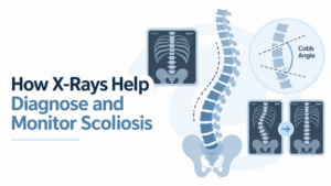
Scoliosis may first be suspected during a physical exam. Uneven shoulders or hips, a shift in posture, or a more prominent rib area when bending forward can all prompt a closer look.
X-rays allow doctors to see the spinal curve directly and measure its size. When scoliosis needs to be monitored, later images can also be compared with earlier studies to see whether the curve has changed.
How Is Scoliosis Diagnosed?
A physical examination usually comes first. Your healthcare provider may assess your posture, shoulder and hip alignment, and the appearance of your back while you stand and bend forward.
If scoliosis is suspected, X-rays of the spine may be ordered. The Scoliosis Research Society’s guide to diagnosing scoliosis explains that standing spinal radiographs allow clinicians to view the curve and measure it in degrees.
This helps confirm whether scoliosis is present and provides a baseline for future comparison when follow-up is needed.
Why Are Scoliosis X-Rays Often Taken While Standing?
Standing images show the spine while it is supporting the body’s weight, which is important when evaluating alignment and curvature.
For an initial scoliosis evaluation, imaging commonly includes upright views of the entire spine from the front or back and from the side. These views allow clinicians to assess the curve in relation to the rest of the spine and pelvis.
Additional views may be ordered in certain situations, so the exact examination can vary from one patient to another.
What Is the Cobb Angle?
The Cobb angle is the standard measurement used to describe the size of a scoliosis curve.
Using the X-ray, the clinician identifies the vertebrae at the upper and lower ends of the curve and measures the angle between them. This provides a number in degrees that can be used to describe the curvature consistently.
A curve greater than 10 degrees meets the commonly used radiographic definition of scoliosis.
The Cobb angle is only one part of the evaluation. Age, remaining growth, curve pattern, symptoms, and changes seen over time also influence how scoliosis is managed.
What Else Can Doctors See on a Scoliosis X-Ray?
Spinal X-rays can provide information beyond the Cobb angle. Depending on the views obtained, they may help show:
- Where the curve is located
- Whether there is one curve or more than one
- Overall spinal alignment
- The normal front-to-back curves of the spine
- Differences compared with previous images
Side-view X-rays can also help assess thoracic kyphosis and lumbar lordosis, the normal curves seen when the spine is viewed from the side.
How Are X-Rays Used to Monitor Scoliosis?
When scoliosis needs follow-up, later X-rays can be compared with previous studies to determine whether the curvature has remained stable or progressed.
This is particularly relevant for children and adolescents who are still growing because spinal curves may change during periods of growth. Current ACR guidance identifies serial spine radiographs as an important part of follow-up for appropriate patients with scoliosis.
There is no single imaging schedule that applies to everyone. How often X-rays are needed depends on factors such as age, skeletal maturity, curve size, previous changes, and the treatment plan.
Do Repeat Scoliosis X-Rays Use Radiation?
Yes. X-rays use ionizing radiation.
When repeated imaging is necessary, clinicians consider both the value of tracking the spinal curve and the importance of limiting unnecessary exposure. Radiology professionals use techniques intended to obtain the images needed for diagnosis while keeping radiation exposure as low as practical. The RadiologyInfo guide to X-ray radiation safety explains how the benefit of medically necessary imaging is weighed against radiation risk.
This is especially relevant in younger patients who may need imaging at more than one point during growth.
When Might Another Imaging Test Be Needed?
MRI is not routinely necessary for every person with scoliosis.
It may be recommended when a healthcare provider needs to examine the spinal cord and other soft tissues more closely or when certain symptoms, neurological findings, or characteristics of the curve raise additional questions.
Current ACR guidance notes that MRI is generally reserved for situations where there are specific clinical concerns rather than used routinely for all adolescent idiopathic scoliosis.
X-Ray in Rockaway, NJ
ImageCare Radiology provides X-ray in Rockaway, NJ, using digital radiography for a range of bone, joint, and other diagnostic imaging needs. Our Rockaway center uses digital X-ray technology, which captures images electronically for radiologist review and communication with the referring provider.
If your healthcare provider orders spinal X-rays for suspected or known scoliosis, the specific views will be based on the information that needs to be measured or compared.
What Changes Between One X-Ray and the Next?
A scoliosis X-ray records the spinal curve at a particular point in time. When another study is performed later, comparing the two can show whether the Cobb angle or overall alignment has changed.
That comparison can be especially useful during growth, when physicians may be watching for progression. The images, measurements, physical exam, and remaining skeletal growth are considered together when deciding what follow-up is appropriate.