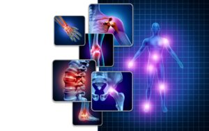
ImageCare Radiology proudly offers arthrography in New Jersey. We provide arthrography procedures to help diagnose and treat joint conditions accurately and effectively. Our team of specialized radiologists uses this diagnostic imaging technique to offer a clear picture of joint health, aiding in the swift and accurate identification of potential issues.
What is Arthrography?
Arthrography is a type of medical imaging used to get a close look at joint structures that may not be visible using standard imaging techniques. This diagnostic procedure is particularly useful for examining joints such as the hips, knees, shoulders, and ankles. During arthrography, a contrast material, or dye, is injected into the joint area to produce highly detailed images. These images can be captured through various technologies including CT scans, MRI scans, and fluoroscopy, which uses X-rays stitched together to create real-time video.
What are the Benefits of Arthrography?
Arthrography is an invaluable tool in the detection of joint issues. It is highly effective in locating tears and damage to the cartilage, ligaments, and tendons of a joint. It reveals parts of the joint that may not be visible with other imaging exams, providing a comprehensive view of the joint’s internal structure. In certain cases, arthrography can be combined with therapeutic procedures such as the injection of local anesthetic medications or steroids into the joint, providing relief from pain or inflammation while also offering valuable diagnostic information.
How Does an Arthrography Procedure Work?
An arthrography procedure at ImageCare Radiology is conducted with the utmost care for patient comfort and safety. Initially, the radiologist administers a local anesthetic to the area around the joint to minimize discomfort. Using image guidance, a needle is then accurately positioned within the joint space, and the contrast material is injected. This contrast material enhances the joint’s internal structures on subsequent CT or MRI scans.
Patients may be asked to move the joint under examination to obtain images from various angles. An arthrogram contrast injection usually takes a few minutes, while the follow-up MRI or CT scan can range from 30 minutes to over an hour, depending on the joint and complexity of the case.
Following the exam, patients may experience some swelling and discomfort. Applying ice and taking over-the-counter analgesics can alleviate these symptoms, which typically subside within 48 hours. We recommend avoiding vigorous exercise for at least 24 hours post-procedure to decrease the risk of joint dislocation. Additionally, minimal activity should be maintained on the affected joint for about 24 hours after the procedure to allow for the injected fluid to dissipate. Vigorous exercise of the joint should be avoided for approximately two weeks to ensure proper healing.
Contact Us
ImageCare Radiology provides accessible and efficient arthrography services. If you require an arthrography procedure or wish to learn more about how it can help diagnose and manage joint-related conditions, please contact us. Our staff is ready to assist you in scheduling an appointment and will support you through every step of the process. Let ImageCare Radiology be your partner in achieving optimal joint health and mobility.