
When Doctors Recommend an Extremity MRI Instead of a CT Scan
If your doctor recommends imaging for an injury or joint problem, you may wonder why one scan is chosen over another. For many patients, terms like MRI and CT scan sound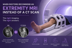 interchangeable, but they actually provide very different types of information.
When evaluating pain or injuries involving the arms, legs, hands, feet, or joints, physicians often select imaging based on what they are trying to see. In some situations, an extremity MRI may provide more useful detail than a CT scan, especially when soft tissues are involved.
Understanding the difference can help patients feel more informed and confident about the imaging process.
What Is an Extremity MRI?
An extremity MRI is a specialized MRI exam focused on areas such as the:
- Shoulder
- Elbow
- Wrist
- Hand
- Hip
- Knee
- Ankle
- Foot
MRI uses magnetic fields and radio waves to create highly detailed images of soft tissues, joints, and surrounding structures without using ionizing radiation.
Unlike standard MRI systems that may require the entire body to enter the scanner, some extremity MRI systems are designed specifically for imaging limbs and joints more comfortably.
How MRI and CT Scans Differ
MRI and CT imaging both play important roles in diagnosis, but they excel at showing different things.
MRI
MRI is particularly effective for evaluating:
- Ligaments
- Tendons
- Muscles
- Cartilage
- Joint inflammation
- Soft tissue injuries
Because of this, MRI is often preferred when doctors suspect injuries that may not involve broken bones.
CT Scan
CT scans use multiple X-ray images to create detailed cross-sectional views of the body.
CT is often better suited for:
- Complex fractures
- Detailed bone imaging
- Certain emergency trauma evaluations
- Situations where imaging speed is especially important
In many cases, CT scans provide excellent detail of bone structures, while MRI offers more detailed views of soft tissues.
When Doctors May Recommend an Extremity MRI
There are several situations where physicians may choose an extremity MRI instead of a CT scan.
Suspected Ligament or Tendon Injuries
MRI is commonly used when doctors suspect injuries such as:
- ACL tears
- Rotator cuff injuries
- Tendon damage
- Sprains involving soft tissue structures
These injuries may not appear clearly on X-rays or CT scans.
Persistent Joint Pain
If a patient has ongoing joint pain without a clear explanation on initial imaging, an MRI may help evaluate cartilage, inflammation, or subtle soft tissue abnormalities.
This is especially common in the:
- Knee
- Shoulder
- Wrist
- Ankle
Sports Injuries
Athletes frequently undergo MRI because sports injuries often involve ligaments, tendons, and cartilage rather than bone alone.
MRI can help physicians better understand the extent of injury and guide treatment planning.
Evaluating Cartilage and Joint Structures
MRI provides detailed images of cartilage and joint surfaces, making it useful for evaluating conditions involving joint degeneration or instability.
When a CT Scan May Still Be the Better Choice
Although MRI offers excellent soft tissue detail, CT scans remain important in many situations.
Doctors may prefer CT imaging when:
- A fracture is suspected after major trauma
- Detailed bone anatomy is needed
- Rapid imaging is required in emergency situations
- MRI is not appropriate for certain patients
The choice depends on the symptoms, medical history, and the structures being evaluated.
What to Expect During an Extremity MRI
During an extremity MRI, the affected joint or limb is positioned within the scanner while detailed images are captured.
Patients are typically asked to remain still during the exam to help ensure clear images. Depending on the area being evaluated, the scan may take anywhere from 20 to 45 minutes.
MRI is painless, though some patients notice sounds from the machine while images are being taken.
Extremity MRI in Sparta, NJ
At ImageCare Radiology, patients seeking an extremity MRI in Sparta, NJ, have access to advanced imaging designed to evaluate joint pain, sports injuries, and soft tissue conditions with precision and detail.
Radiologists work closely with referring providers to help ensure imaging findings support accurate diagnosis and personalized treatment planning.
Choosing the Right Imaging for the Right Problem
MRI and CT scans are both valuable tools, but they answer different medical questions. When soft tissue injuries, joint structures, or persistent pain need closer evaluation, an extremity MRI may provide the detail physicians are looking for.
The most important thing patients should know is that imaging recommendations are made with purpose. Choosing the right exam helps doctors gather the clearest possible information, which ultimately supports better care and more informed treatment decisions.