
Why Is Ultrasound Used to Guide a Biopsy?
How real-time imaging helps doctors locate a target, guide the needle, and collect tissue for testing.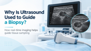
When a suspicious lump, nodule, or other abnormal area needs to be tested, a biopsy can provide information that imaging alone cannot. The procedure involves removing a small sample of cells or tissue so it can be examined in a laboratory.
In some cases, ultrasound helps the physician reach the exact area that needs to be sampled. Because the images appear in real time, the physician can see both the target and the biopsy needle as the procedure is performed.
What Is an Ultrasound-Guided Biopsy?
An ultrasound-guided biopsy combines diagnostic ultrasound with needle biopsy.
Ultrasound uses sound waves to create live images of structures inside the body. Once the area being sampled is visible, the physician can use those images to plan the needle path and guide the needle toward the target.
The RadiologyInfo overview of image-guided biopsy explains that ultrasound is one of several imaging methods physicians may use to guide a needle to an abnormal area for tissue sampling.
Why Is Real-Time Imaging Helpful?
Unlike a still image, ultrasound allows the physician to watch the needle move on the screen during the procedure.
That can help with:
- Locating the area that needs to be sampled
- Choosing an appropriate path for the needle
- Following the needle as it approaches the target
- Seeing nearby structures while the sample is collected
Ultrasound also uses sound waves rather than ionizing radiation.
Whether it is the best guidance method depends on where the abnormality is located and how clearly it can be seen on ultrasound.
What Is the Difference Between Fine-Needle Aspiration and Core Biopsy?
Ultrasound guidance can be used with different types of needle biopsy.
A fine-needle aspiration, or FNA, uses a thin needle to remove cells or fluid from an area of concern.
A core needle biopsy uses a slightly larger needle to collect small pieces of tissue. Preserving the tissue structure can provide the laboratory with different information than a sample made up primarily of individual cells.
Your healthcare team chooses the technique based on the area being evaluated and the information needed from the sample.
Can Ultrasound Tell Whether a Mass Is Cancer?
Ultrasound can provide useful information about the appearance of a lump, nodule, or other abnormality, but the image itself may not be able to determine exactly what the tissue represents.
That is where the biopsy becomes important.
Once the sample is collected, it is sent to a laboratory, where a pathologist examines the cells or tissue. The pathology findings help determine whether the sample is benign, cancerous, or shows another type of condition.
Why Would CT or Another Imaging Method Be Used Instead?
Ultrasound is well suited for abnormalities that can be clearly visualized with sound-wave imaging, particularly in accessible soft tissues and organs.
Other areas may be easier to reach using a different form of guidance. CT, for example, can provide detailed cross-sectional images when the target is deeper in the body or difficult to see on ultrasound. Certain breast findings may instead be sampled using mammography-based stereotactic guidance or MRI.
The imaging method is chosen according to the location of the abnormality and which approach allows the physician to guide the needle accurately.
Does an Ultrasound-Guided Biopsy Require Special Preparation?
Preparation varies according to the body part being biopsied, the type of needle used, and other details of the procedure.
You may receive instructions about medications, eating or drinking, transportation, or activity afterward. Follow the directions provided for your specific biopsy rather than relying on general instructions online.
In particular, do not stop blood thinners, aspirin, or other medications unless the healthcare professional managing your procedure tells you to do so.
Ultrasound in Wayne, NJ
ImageCare Radiology provides ultrasound in Wayne, NJ, for a range of diagnostic needs. Ultrasound can also be used as an imaging-guidance tool during certain procedures when a physician needs a real-time view of the area being targeted.
The type of ultrasound or image-guided procedure your provider recommends depends on what needs to be evaluated.
What Happens to the Biopsy Sample?
After tissue or cells are collected, the sample is sent to a pathology laboratory for examination. The physician who ordered or performed the biopsy then uses the pathology results along with your imaging and other medical information to determine what comes next.
If you have an ultrasound-guided biopsy scheduled, knowing why ultrasound is being used can make the procedure easier to understand. Its role is to help the physician see the target and guide the needle accurately, while the tissue sample provides the information needed for diagnosis.