World Scleroderma Day: How Musculoskeletal Imaging Helps Evaluate Joint and Tissue Changes
Each year on June 29th, World Scleroderma Day raises awareness about a rare autoimmune condition that affects connective tissues throughout the body. While many people associate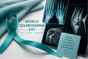 scleroderma primarily with changes to the skin, the condition can also affect the joints, muscles, blood vessels, and internal organs.
For some patients, symptoms involving movement and mobility become an important part of daily life. Joint stiffness, hand pain, muscle weakness, and reduced flexibility can all impact comfort and function over time.
Because these changes often develop gradually and affect structures beneath the surface, musculoskeletal imaging can help physicians better evaluate how the condition may be affecting the body.
What Is Scleroderma?
Scleroderma is a chronic autoimmune disease that causes the body to produce excess collagen, a protein found in connective tissues. This can lead to thickening and tightening of the skin, along with inflammation and tissue changes in other parts of the body.
There are different forms of scleroderma, and symptoms can vary significantly from person to person.
Some individuals experience primarily skin-related symptoms, while others may develop issues involving:
- Joints
- Muscles
- Tendons
- Blood vessels
- Internal organs
Because the condition can affect multiple systems, evaluation often involves a team of healthcare providers.
How Scleroderma Can Affect the Musculoskeletal System
Scleroderma may impact the musculoskeletal system in several ways. Inflammation, tissue thickening, and changes in connective tissue can affect movement and flexibility over time.
Common musculoskeletal symptoms may include:
- Joint pain or stiffness
- Swelling in the hands or fingers
- Muscle weakness
- Reduced range of motion
- Tightening around joints
Some patients may also develop calcinosis, a condition where calcium deposits form beneath the skin or around soft tissues.
These symptoms can make everyday tasks more difficult and may gradually affect mobility if not properly monitored.
Why Imaging May Be Recommended
Physical exams and symptom discussions remain essential in evaluating scleroderma, but imaging can provide additional insight into what may be happening beneath the surface.
Musculoskeletal imaging helps physicians assess:
- Joint inflammation
- Bone and joint changes
- Soft tissue involvement
- Calcified deposits
- Structural changes affecting mobility
Imaging may also help doctors monitor how symptoms are changing over time or evaluate complications related to the condition.
Imaging Methods Used in Scleroderma Evaluation
Different imaging techniques may be used depending on the symptoms being evaluated.
X-Ray
X-rays can help identify changes involving bones and joints, particularly in the hands. They may also detect calcium deposits associated with calcinosis.
MRI
MRI provides detailed images of soft tissues, muscles, tendons, and joints. This type of imaging may help evaluate inflammation or deeper tissue involvement.
Ultrasound
Ultrasound may be used to assess joints, tendons, and soft tissue structures in certain situations. It can also help evaluate inflammation in a noninvasive way.
Each imaging method offers different information, helping physicians build a more complete picture of how the condition may be affecting the body.
Monitoring Changes Over Time
Scleroderma symptoms can change gradually, which is why ongoing monitoring is often an important part of care.
Imaging may help physicians:
- Track progression of joint or tissue changes
- Evaluate new or worsening symptoms
- Support treatment planning
- Monitor mobility-related complications
Because no two cases are exactly alike, imaging is typically used alongside clinical evaluation and other medical testing.
Musculoskeletal Imaging at ImageCare Radiology
At ImageCare Radiology, advanced musculoskeletal imaging helps physicians evaluate conditions affecting the joints, muscles, bones, and connective tissues. Detailed imaging allows radiologists to assess structural changes and provide information that supports diagnosis and ongoing care.
Radiologists work closely with referring providers to ensure imaging findings contribute meaningfully to each patient’s overall treatment plan.
Raising Awareness During World Scleroderma Day
World Scleroderma Day helps bring attention to a condition that is often misunderstood and difficult to recognize early. Increased awareness can encourage earlier evaluation, better symptom management, and improved support for patients living with the disease.
While imaging cannot diagnose scleroderma on its own, musculoskeletal imaging plays an important role in helping physicians understand how the condition may be affecting joints, muscles, and connective tissues over time.
For patients navigating complex symptoms, that added insight can help support more informed and personalized care.