World Hemophilia Day: How Imaging Helps Protect Joint Health
Every year on April 17, World Hemophilia Day raises awareness about a group of inherited bleeding disorders that affect the body’s ability to form blood clots. While hemophilia is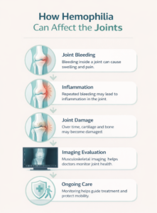 often associated with bleeding episodes, one of the most significant long-term concerns involves joint health.
Repeated bleeding into joints can gradually damage cartilage, bone, and surrounding tissues. Over time, this can lead to pain, stiffness, and reduced mobility. For many patients, protecting joint function becomes an important part of managing hemophilia.
This is where musculoskeletal imaging can play a valuable role. Imaging helps physicians evaluate joint structures, monitor changes over time, and better understand how hemophilia may be affecting the body.
Understanding Hemophilia
Hemophilia is a genetic bleeding disorder caused by a deficiency in certain clotting factors that help the blood form clots. Without these factors, bleeding can last longer than normal after an injury.
In some cases, bleeding can occur inside the body, particularly within joints and muscles. These episodes are sometimes referred to as joint bleeds or hemarthrosis.
The joints most commonly affected include:
- Knees
- Ankles
- Elbows
Because joints experience frequent movement and stress, bleeding in these areas can cause inflammation and damage if it occurs repeatedly.
Why Joint Bleeding Happens in Hemophilia
Joint bleeding occurs when small blood vessels inside the joint become injured or stressed. In individuals without hemophilia, the body quickly forms a clot to stop the bleeding. For people with hemophilia, the clotting process may take longer.
When blood enters the joint space, it can lead to:
- Swelling
- Pain or warmth in the joint
- Reduced range of motion
- Long-term cartilage damage if episodes repeat
Repeated bleeding over time may lead to a condition known as hemophilic arthropathy, a form of joint disease caused by chronic bleeding.
Early evaluation and monitoring are important to help protect joint function.
How Musculoskeletal Imaging Supports Hemophilia Care
Musculoskeletal imaging focuses on evaluating bones, joints, and soft tissues. For individuals living with hemophilia, imaging helps doctors better understand how joints are affected by bleeding episodes.
Imaging can help physicians:
- Evaluate joint inflammation or swelling
- Detect early changes in cartilage or bone
- Monitor joint health over time
- Identify complications related to repeated bleeding
By providing a clearer view of what is happening inside the joint, imaging can support treatment decisions and help guide long-term care.
Imaging Methods Used to Evaluate Joint Health
Different imaging techniques may be used depending on the situation and the joint being examined.
Ultrasound
Ultrasound can help physicians identify fluid or bleeding within the joint. Because it uses sound waves rather than radiation, it is often used for evaluating joint swelling and soft tissues.
MRI (Magnetic Resonance Imaging)
MRI provides detailed images of soft tissues, cartilage, and joint structures. This imaging method can help detect early joint changes that may not yet be visible on other imaging studies.
X-ray
X-rays can help evaluate bone structure and joint alignment. They may be used to identify long-term joint changes associated with chronic bleeding.
Each imaging method provides a different perspective, allowing physicians to gather a more complete understanding of joint health.
Monitoring Joint Health Over Time
For individuals with hemophilia, managing joint health is often an ongoing process. Imaging studies may be used periodically to help physicians monitor how joints are changing and determine whether treatment approaches are working effectively.
Monitoring can help doctors:
- Detect joint changes early
- Adjust treatment strategies when needed
- Support mobility and long-term joint function
With careful medical management and monitoring, many people with hemophilia are able to maintain active and fulfilling lives.
Musculoskeletal Imaging at ImageCare Radiology
At ImageCare Radiology, musculoskeletal imaging helps physicians evaluate conditions affecting bones, joints, and surrounding tissues. Advanced imaging technologies allow radiologists to carefully assess joint structures and provide insights that support diagnosis and treatment planning.
Radiologists work closely with referring providers to ensure imaging results contribute meaningfully to patient care.
Raising Awareness During World Hemophilia Day
World Hemophilia Day serves as a reminder of the importance of awareness, education, and access to specialized care for individuals living with bleeding disorders.
While hemophilia affects the body’s ability to clot blood, modern medical care, including musculoskeletal imaging, helps physicians better understand and monitor how the condition affects joint health.
By improving awareness and encouraging proactive care, healthcare providers and families can work together to help protect mobility, reduce complications, and support long-term quality of life.