How Neuroradiology Supports Neurosurgical Care
Neurosurgery Awareness Month is observed each August and led by the American Association of Neurological Surgeons. The observance highlights the role of neurosurgery in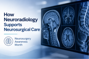 advancing care for conditions that affect the brain, spine, and nervous system.
For patients, this month is also a helpful opportunity to better understand the care team behind complex neurological and spine conditions. Neurosurgical care often involves more than one provider, and imaging can play an important role in helping physicians evaluate what is happening inside the body.
That is where neuroradiology comes in. Neuroradiology is a subspecialty of radiology focused on imaging and diagnosing conditions involving the brain, spine, head, and neck.
What Is Neuroradiology?
Neuroradiology focuses on detailed imaging of the central and peripheral nervous system, including the brain, spine, spinal cord, head, neck, and nerves. According to the American Society of Neuroradiology, neuroradiologists interpret imaging studies such as MRI, CT scans, X-rays, and angiographic studies involving the brain and spine.
In simpler terms, neuroradiology helps doctors get a clearer view of areas that are often difficult to evaluate through symptoms or a physical exam alone.
Neuroradiology may be used to help assess conditions involving:
- The brain
- The spine and spinal cord
- The head and neck
- The nerves
- Blood vessels in the brain or spine
- Certain injuries, abnormalities, or changes over time
It does not replace a neurosurgeon’s evaluation. Instead, it provides important visual information that can help guide the next step in care.
Callout: Neuroradiology Is About Clarity
Brain and spine symptoms can be complex. Neuroradiology helps physicians see details that may not be obvious from symptoms alone, giving the care team more information to work with.
How Neuroradiology Connects to Neurosurgery Awareness Month
Neurosurgery Awareness Month recognizes the work of neurosurgeons and the impact of modern neurosurgical care. But before a neurosurgeon can recommend the right treatment approach, they often need detailed imaging.
Neuroradiology supports that process by helping physicians better understand the location, size, structure, and possible cause of a neurological or spine concern.
For example, imaging may help a provider evaluate:
- Back or neck pain that may involve the spine
- Headaches or neurological symptoms that require further investigation
- Possible brain or spinal abnormalities
- Injury after trauma
- Tumors or masses
- Stroke-related concerns
- Changes after surgery or treatment
The imaging findings can help doctors decide whether a patient needs monitoring, medication, physical therapy, additional testing, specialist care, or a surgical consultation.
Common Imaging Tests Used in Neuroradiology
Different imaging tests answer different clinical questions. Your provider will recommend the test that best fits your symptoms, medical history, and the area being evaluated.
Common neuroradiology imaging tools include:
- MRI: Often used to create detailed images of soft tissues, the brain, spine, spinal cord, discs, nerves, and surrounding structures.
- CT scan: Provides detailed cross-sectional images and may be used when speed, bone detail, trauma evaluation, or certain urgent concerns are important.
- X-ray: May be used to evaluate bones, alignment, or certain spine-related concerns.
- Angiography or vascular imaging: Helps evaluate blood vessels in the brain, head, neck, or spine when vascular detail is needed.
RadiologyInfo, a patient resource from the Radiological Society of North America and the American College of Radiology, explains that imaging resources help patients understand how exams such as X-ray, CT, MRI, and ultrasound are performed and how to prepare for them.
Why MRI Is Often Important for Brain and Spine Imaging
MRI is commonly used in neuroradiology because it provides detailed views of soft tissue without using ionizing radiation. This can be especially useful when evaluating the brain, spinal cord, discs, nerves, and other soft-tissue structures.
A spine MRI may help doctors look at the spinal cord, nerve roots, discs, and surrounding tissues. RadiologyInfo notes that spine MRI can provide detailed pictures that help physicians evaluate a variety of spine-related concerns.
MRI may be recommended when a provider needs to better understand symptoms such as:
- Radiating pain
- Numbness or tingling
- Weakness
- Persistent neck or back pain
- Changes in coordination or balance
- Symptoms that suggest nerve involvement
The goal is not simply to “find something.” The goal is to gather information that helps your physician understand what may be contributing to your symptoms.
Callout: Imaging Helps Guide the Next Step
A scan does not make a treatment decision on its own. Your doctor reviews imaging alongside your symptoms, exam findings, health history, and other test results.
How Imaging Can Support Care Before and After Treatment
Neuroradiology can be helpful at several points in a patient’s care journey. Before treatment, imaging may help physicians:
- Identify the area of concern
- Understand whether nerves, discs, bones, blood vessels, or soft tissues are involved
- Determine whether additional testing is needed
- Decide whether conservative care or specialist referral may be appropriate
- Plan for possible procedures or surgery when needed
After treatment, imaging may help doctors:
- Monitor healing
- Compare changes over time
- Evaluate new or returning symptoms
- Check the results of a procedure or surgery
- Guide ongoing follow-up care
This is one reason neuroradiology is so closely connected to neurosurgical care. Clear imaging helps create a clearer path forward.
What Patients Should Know Before a Neuroimaging Exam
If your doctor recommends brain, spine, or nerve-related imaging, it is natural to have questions. The type of exam you receive will depend on what your provider needs to see.
Before your appointment, it may help to ask:
- What area of the body is being imaged?
- Why is this test being recommended?
- Should I bring prior imaging or reports?
- Do I need to avoid eating or drinking beforehand?
- Will contrast be used?
- How will I receive my results?
You should also tell the imaging team if you have implanted medical devices, prior surgeries, kidney concerns, allergies, or difficulty lying still. These details can help the team prepare for your exam safely and comfortably.
A Clearer View of Brain and Spine Health
Neurosurgery Awareness Month is a reminder of how important brain, spine, and nervous system care can be. Neuroradiology supports that care by giving physicians detailed images that can help with diagnosis, treatment planning, and follow-up.
For patients, that means imaging can provide answers when symptoms are unclear, help doctors monitor changes, and support more informed conversations about next steps. Whether your provider recommends MRI, CT, X-ray, or another imaging exam, the purpose is the same: to better understand what is happening and guide care with confidence.
If your doctor has recommended neuroimaging, ImageCare Radiology offers patient-focused diagnostic imaging services to help support your next step in care.