How Imaging Helps Stage and Monitor Lung Cancer
How CT, PET/CT, and other scans help doctors understand the extent of lung cancer and follow changes during treatment.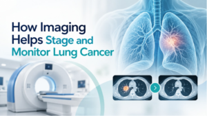
Lung Cancer Awareness Month in November often puts a well-deserved focus on screening and early detection. Imaging also remains important after a suspicious finding or confirmed lung cancer diagnosis.
At that point, doctors may need to know the size and location of a tumor, whether nearby lymph nodes are involved, whether cancer has reached another part of the body, or how the disease is responding to treatment. Different imaging tests can help answer those questions at different stages of care.
What Can a CT Scan Show About Lung Cancer?
CT creates detailed cross-sectional images of the chest. When doctors are evaluating a suspicious lung finding, these images can show its size, shape, and location, along with enlarged lymph nodes or other abnormalities that may need a closer look.
The American Cancer Society’s guide to lung cancer testing explains that CT is also used to look for possible spread and to evaluate changes during treatment.
A suspicious area on CT does not necessarily mean someone has lung cancer. When cancer is suspected, examining cells or tissue in a laboratory is generally needed to confirm the diagnosis and determine the type of cancer.
How Does Imaging Help Stage Lung Cancer?
After lung cancer is diagnosed, staging describes how much cancer is present and where it is in the body.
For non-small cell lung cancer, doctors consider the size and extent of the primary tumor, whether nearby lymph nodes contain cancer, and whether it has spread to distant areas.
Imaging can help evaluate:
- The original tumor in the lung
- Nearby tissues and structures
- Lymph nodes
- Areas elsewhere in the body where cancer may have spread
The exams used for staging vary with the type of lung cancer and what doctors already know from earlier tests. Current ACR-based guidance includes chest CT, PET/CT, and brain MRI among the studies that may be appropriate when staging lung cancer.
Why Might a Doctor Order a PET/CT?
PET/CT combines two kinds of information in one examination.
The CT portion shows detailed anatomy. PET uses a radioactive tracer, often a form of glucose, to show areas of increased metabolic activity. When the images are viewed together, doctors can see where an area of increased activity is located in the body.
This can be particularly useful when evaluating whether lung cancer may have reached lymph nodes or other areas. The American Cancer Society’s explanation of PET imaging also notes that PET can be used for cancer staging and, in some situations, to evaluate response to treatment.
An area that lights up on PET is not automatically cancer. Infection and inflammation can also cause increased tracer uptake, so doctors interpret those findings along with the rest of the patient’s imaging and medical information.
When Is MRI Used?
MRI is not usually the primary test for examining a lung tumor itself, but it can become important when doctors need detailed images of certain other areas.
For example, brain MRI may be used to look for lung cancer that has spread to the brain. MRI can also be helpful for selected questions involving other soft tissues.
Which exams are needed depends on the type and stage of the cancer as well as the patient’s symptoms and previous test results.
How Does Imaging Help Monitor Treatment?
Follow-up imaging allows doctors to compare findings over time. CT may show whether a tumor has changed in size or appearance. PET can sometimes provide additional information about changes in metabolic activity. Imaging may also be used after treatment to look for possible recurrence.
There is no universal scan schedule for every person with lung cancer. The type and timing of imaging depend on the cancer, treatment plan, and clinical questions that need to be answered.
Does an Abnormal Scan Always Mean Lung Cancer?
No. Lung nodules and other abnormalities can have noncancerous causes.
Imaging helps doctors characterize a finding and decide what, if anything, should happen next. Depending on the circumstances, that could mean another scan, follow-up imaging after a period of time, or a biopsy.
This is also why patients should be cautious about trying to interpret lung CT or PET images on their own. A radiologist considers the appearance of a finding along with its location, previous images, and other clinical information.
The Role of Oncology Imaging
Imaging can serve different purposes as someone moves through cancer evaluation and care. CT may provide detailed anatomical information, PET/CT can help evaluate areas of metabolic activity, and MRI may answer questions about areas such as the brain.
ImageCare Radiology offers oncology imaging services that include CT, MRI, and PET/CT, among other imaging methods used in cancer evaluation and follow-up.
If several scans are recommended, it is reasonable to ask what your care team hopes to learn from each one. Knowing whether a test is being used to evaluate the original tumor, determine whether cancer has spread, or monitor a change over time can make the reason for the exam much clearer.
What Imaging Can Tell You Over Time
Imaging provides information at several points in lung cancer care, but each exam has a specific purpose. A scan used for staging may answer a different question from one performed months later during treatment or follow-up.
For patients, understanding that purpose can make a series of unfamiliar tests easier to follow. For the care team, those images provide another source of information for deciding what should happen next.